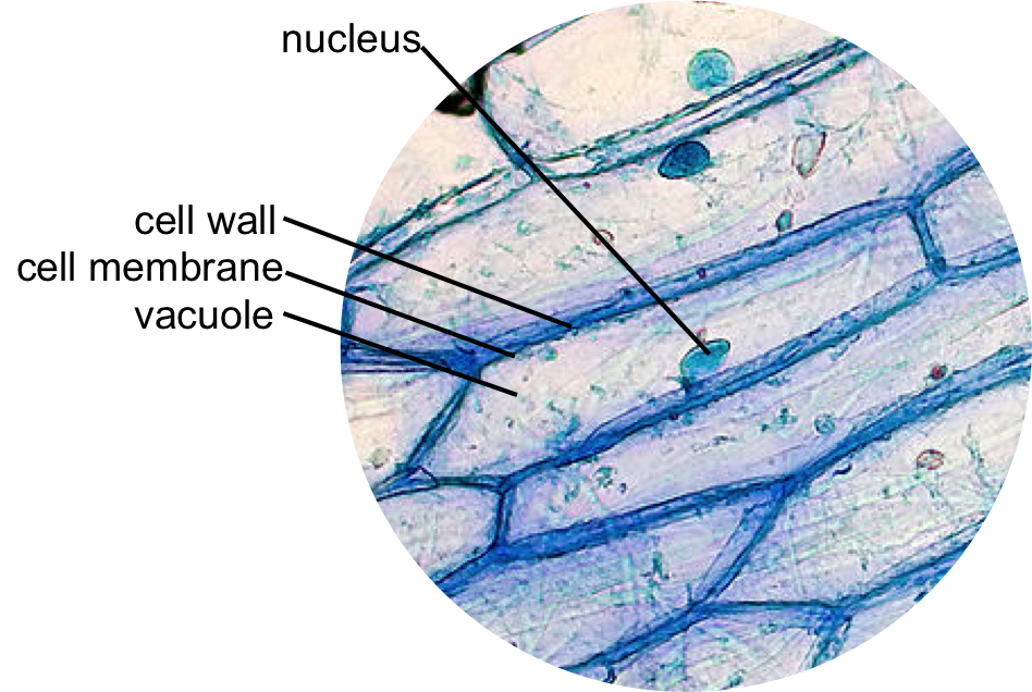
animal cell under light microscope
All eukaryotic cells have Nucleus few cells such as the mammalian RBCs may do not have. A typical animal cell is 1020 μm in diameter which is about one-fifth the size of the smallest particle visible to the naked eye.

Onion Epidermis With Large Cells Under Light Microscope Clear Epidermal Cells O Ad Microscope Light Epiderma Microscopic Cells Dna Project Plant Cell
Since most cells are between 1 and 100 μm in diameter they can be observed by light microscopy as can some of.

. Make a wet or dry mount with a coverslip. Make a wet mount slide. Once slides have been prepared they can be examined under a microscope.
The cells youll be looking at in this activity were photographed with a light microsope. Onions are multicellular plant organisms which basically means that they are made up of many cells that are uniform in. Animal cell under the microscope.
Novel Coronavirus SARS-CoV-2 Under the Microscope The National Institute of Allergy and Infectious Diseases Rocky Mountain Laboratories NIAID-RML located in Hamilton Montana was able to capture images of the novel coronavirus SARS-CoV-2 previously known as 2019-nCoV on its scanning electron microscope and transmission electron microscopes. The seasoned team soon confirmed the death was a murder but no footprints no fingerprints no weapons were founda few strands of hair caught in the dead womans broken fingernails were the only evidence the killer left behind. Histology also known as microscopic anatomy or microanatomy is the branch of biology which studies the microscopic anatomy of biological tissues.
Iodine crystal violet and methylene blue are examples of simple stains. When viewed under the light microscope Euglena appear as elongated unicellular organisms that are rapidly moving across the field surface. The discovery of the cell would not have been possible if not for advancements to the microscope.
Pieces of the tumor are then examined by a veterinary pathologist under the microscope. Under the microscope animal cells appear different based on the type of the cell. His microscope used three lenses and a stage light which lit up and enlarged the specimens.
In order to examine cells in the tip of an onion root a thin slice of the root is placed onto a microscope slide and stained so the chromosomes will be visible. Observing Onion Cells under a Microscope. Histology is the microscopic counterpart to gross anatomy which looks at larger structures visible without a microscope.
A nucleus or a cell wall can be seen more clearly by using different stains. The light microscope remains a basic tool of cell biologists with technical improvements allowing the visualization of ever-increasing details of cell structure. The goals for this lesson are to.
These regions of growth are good for studying the cell cycle because at any given time you can find cells that are undergoing mitosis. There are two microscope lesson activities in this blog for you to see the nuclei in animal cells and plant cells. An experienced forensic scientist can easily tell if the hair specimen is from humans or from animals.
Even though plant cells are eukaryotic the difference can be easily identified as the animal cells lack. Animal cell nucleus function plays the most important role for the cell. We can determine the number of chromosomes and chromatids per chromosome for each stage in mitosis.
Most plant and animal cells are only visible under a light microscope with dimensions between 1 and 100 micrometres. I thought it would be helpful to share how I help students to see an example of a plant cell. Observe an onion cell under the microscope.
Two cells will be observed one from the skin of an onion and the other from a common aquarium water plant anacharis. This is a solid microscope aimed mostly at high school level education and comes with a small microscope camera all for under 1000. It comprises other cellular structures and organelles which helps in carrying out some specific functions required for the proper functioning of the cell.
By comparing different. In some cases results from FNA may not be entirely clear and biopsy may be necessary. The cell from the Latin word cellula meaning small room is the basic structural and functional unit of lifeEvery cell consists of a cytoplasm enclosed within a membrane which contains many biomolecules such as proteins and nucleic acids.
We found that although it is a nice microscope for high schoolers who use it maybe half an hour a day every few days the kind of detail needed by the hair analysis professionals requires stronger illumination and a higher-octane camera. Chromosomes are usually visible under light microscope. What do onion cells look like under the microscope.
A typical animal cell is 1020 μm in diameter which is about one-fifth the size of the smallest particle visible to the naked eye. Much later in 1831 Robert Brown an Englishman observed that all cells had a centrally positioned body which he termed. In biological terms an animal cell is a typical eukaryotic cell with a membrane-bound nucleus with DNA present inside the nucleus.
This is called histopathology. What does it mean to draw a cell to scale 5. List 2 organelles that were NOT visible but could be found in cells if you had a microscope with a better magnification.
Confocal laser scanning Microscope It uses several mirrors that scan along the X and Y axes on the specimen by scanning and descanning and the image passes through a pinhole into the detector. This discovery proposed as the cell doctrine by Schleiden and. To use a light microscope to examine animal or plant.
A biopsy is a surgical excision of a piece of the tumor. Round is the most common shape of the nucleus but depending on the type of cell. However the internal structure and organelles are more or less similar.
In 1672 Leeuwenhoek observed bacteria sperms and red blood corpuscles all of which were cells. Interested in learning more about the microscopic world scientist Robert Hooke improved the design of the existing compound microscope in 1665. Although one may divide microscopic anatomy into organology the study of organs histology the study of.
There are various number of nuclei either they are single nucleus uni-nucleate two nuclei bi-nucleate or multi-nucleate. Plant Cell Lab Makeup Purpose. Contemporary light microscopes are able to magnify objects up to about a thousand times.
Types of Confocal Microscope. Keeping in mind that the. A veterinary pathologist then examines the slide under a microscope.
1665 observed a piece of cork under the microscope and found it to be made of small compartments which he called cells Latin cell small room. It is easier to see nuclei under a light microscope with staining such as methylene blue. The light microscope used in the lab is not powerful enough to view other organelles in the cheek cell.
They usually study the hairs scale pattern its color and the appearance of the medulla. Aims of the experiment. The morphology under a microscope.
Investigating cells with a light microscope. Chromatin in the nucleus begins to condense and becomes visible under the light microscope as chromosomes each with two chromatids that are held together at the centromere. How to see the cell nucleus under a microscope.
Get Forensic with Hair Analysis Life was long gone from the cold bloody corpse when the crime scene investigators arrived. Microscope cell staining is a technique used to improve the visibility of cells and cell parts under a microscope. Studying cell tissues from an onion peel is a great exercise in using light microscopes and learning about plant cells since onion cells are highly visible under a microscope especially when stained correctly.
Although this is not the case with all it is the most common. What parts of the cell were visible. Students will observe plant cells using a light microscope.
One of the easiest labs in cell biology is observing onion cells under a microscope. It was not until good light microscopes became available in the early part of the nineteenth century that all plant and animal tissues were discovered to be aggregates of individual cells. One thing that students will notice as soon as they begin to observe the organism is that it has a blunt rounded end portion and a pointed end this gives them a tear-drop shape.
Spinning disk also known as the Nipkow disk is a type of confocal microscope that uses several movable apertures pinholes. Forensic scientists view hair under a microscope to collect evidence based on morphology.

Year 11 Bio Key Points Cell Organelles And Their Function Cell Organelles Animal Cell Organelles

Epidermal Onion Cells Under A Microscope Plant Cells Appear Polygonal From The Plant Cell Diagram Cell Diagram Plant Cell

Structure Of Animal Cell And Plant Cell Under Microscope Diagrams Animal Cell Cell Diagram Plant And Animal Cells

List Of Cell Organelles Their Functions Animal Cell Plant And Animal Cells Plant Cell

Cheek Cells Things Under A Microscope Cell Cheek

Cell 8 Pictures Of Plant Cells Under A Microscope Plant Cell Structure Under Microscope Plant And Animal Cells Plant Cell Structure Plant Cell Picture

Animal Cell Organelles Sauna Design

Animal Cell Structure Cell Organelles Organelles

Cell Structure Human Cell Diagram Animal Cell Drawing Cell Diagram